

Hírek
Szavazás
Live Blood Analysis
Live Blood Analysis
INTRODUCTION
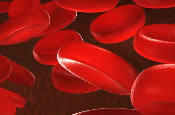
Live Blood Analysis uses a drop of live blood from the patients finger that has not been killed by staining, and viewed under a special microscope using a darkfield condenser. This enables the blood sample to be illuminated from the sides, making the various components phosphoresce behind a dark background. This makes it possible to see very small particles, smaller than a cell that would not normally be visible under a normal light microscope. All the living components of the blood are seen clearly, and can be viewed by the patient and therapist using a video camera and a dedicated monitor.
The examination of live blood is valuable for the early detection of serious health conditions. It is possible to see at what stage of pathological development the body is in, simply from using one drop
of blood.
Because the precursors to serious health imbalances may be
observed in the state of ever-present floras found in the blood,
health imbalances may be averted by reading these early warning signs and making the necessary changes that will allow one to rebalance the physiology. These markers are also applicable in the course of tracking the progression and reversal of degenerative conditions that may already be in motion.
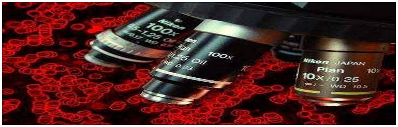
DARKFIELD MICROSCOPY
The observations of these floras are made using what is known as
a Darkfield microscope. When utilizing todays conventional approaches to analyze blood, the standard methods are to use
either stains, which make certain factors in the blood visible which would not be otherwise visible, or the electron microscope, which provides ultrahigh magnifications. The limitation of both of the preceding approaches is that the blood is effectively killed through
the processes utilized in observation. In a Darkfield, the blood is stained with light frequencies, which allow the blood to remain alive and active, giving many otherwise unobtainable clues as to the relative health of the blood, thereby, the whole organism.
We have entered into a new era of scientific discovery. As the
horse gave way to the horseless carriage, so must science now accommodate a new understanding of the body as a whole.
Edgar Cayce, the seer of Virginia Beach, predicted in the 1930s that
in the future, a persons state of health would be determined by the evaluation of one drop of blood. This time has arrived!
Through the advent of technological advances in microscopy, new discoveries have been proven by such leading researchers as Royal Rife, Gaston Naessans, Dr. Gunther Enderlein, Dr. Majid Ali, M.D. and many others. In the rapidly emerging field of what is known as live
cell analysis, an understanding of biology as a holistic science has emerged. Health imbalances in the body may be averted by observing the state of ever-present floras found in the blood and by correcting the milieu that allows the floras to remain in their regulatory forms
or to move into pathogenicity. Or conversely, to be reduced from pathogenic forms back down to regulators. These observations are made in what is known as a darkfield. 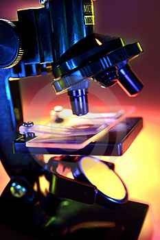
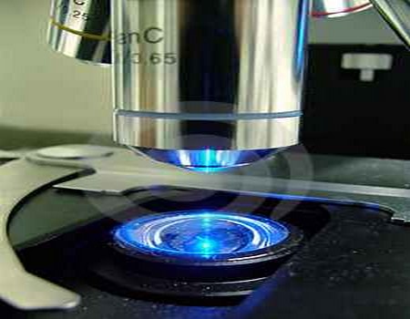
DARK-FIELD SYSTEMS
Under dark-field examination, the various materials making up the structure of the cell or microorganism under view cause it to appear
to glow and emit its own light. Since fine structures often cannot be seen when they appear in front of a bright background, illuminating them from the side and viewing them on a black background lights them up and provides contrast and super fine resolution when plan achromat oil immersion iris diaphragm objectives are used. The background is black because the direct or transmitted light from the condenser is passing around the objective and not directly into it.
The only light that is seen is light that is reflected off of the sides
and surfaces of the particles. What are seen are illuminated particles on a black background.
This provides a very clear view of the minutest forms, as the eyes potential to differentiate is not overwhelmed by background light
and light passing through the sample. Viewing blood in a dark-field is a very useful adjunct when evaluating and addressing the traumatic factors in the total life picture of the individual. The advanced phases of the life-cycle of the colloidal microorganisms are at the very
deepest level of causes of blood imbalance and other organism aberrations
WHAT CAN BE DETERMINED FROM LIVE BLOOD ANALYSIS?
Depending on the irregularities found in the blood, there are a wide variety of different conditions that can be determined. The following are just a few examples:
· Indication of low immune status
· Liver and spleen stress
· Vitamin and mineral deficiencies
· Hormonal imbalances
· Fungal infections
· Parasite infestation
· Digestive problems
· Atherosclerotic predisposition
· Heavy metal toxicity
· Predisposition to cancer or other degenerative diseases

IS BLOOD STERILE?
For a long time many researchers and doctors believed, and many
still do, that blood is sterile. Professor Dr. Gunter Enderlein after numerous years of research, proved otherwise. He showed using darkfield microscopy, that the serum of all people and warm-blooded animals are alive with many moving particles. He called these particles ENDOBIONTS (from the Greek "endon" = internal and
"bios" = life). When you first look at a live blood sample under a microscope or on a monitor you cannot fail to see a multitude of moving, living particles. These are the Endobionts, and are an important part of health.
There are a wide variety of different Endobionts in the blood. In
fact, from the simplest apathogenic (non-disease forming) PROTIT, they
can change forms into pathological (disease causing) species, depending on how much the pH (acidity-alkalinity) of the blood is changed. The higher the acidity, the more pathogenicity, and the
more likelihood of developing a chronic, degenerative disease
process over time. The wonderful world of Live Blood Analysis can enable you to see the different forms in the blood, and to therefore determine how far ahead you are in the disease process.
Prof. Enderlein discovered that these Endobionts change into
different forms as the internal milieu changes. The more the metamorphosis (different changes), the greater the probability of disease. The pH of our blood is determined by the food we eat,
and the nutrients we take in daily, as well as other factors such as stress and pollutants. Fast and refined foods and concentrated protein foods all lead to acidic blood which triggers the Endobiont
to change to more pathogenic forms. All microbes partake in a
natural developmental cycle, that begins with the PRIMITIVE PHASE which is microscopically invisible; this changes into the BACTERIAL PHASE; and finally culminates in the FUNGAL PHASE, which is the
most pathogenic stage. This is the theory of PLEOMORPHISM (many forms), as opposed to monomorphism (one form).
BABY - START - PROGRAM
Térítésmentes
Egészségmegőrző Tanácsadás
Budapest
Bijó Öngyógyító
központ

.
BABY - START
program

Gyermeket szeretne?
Tudatosan készül a gyermekvállalásra?
A legjobb helyen jár…
.
infó:
20/9464-249
Naptár
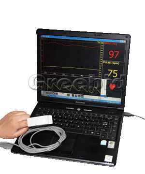
MINŐSÉGI VÉRELEMZÉS
FÉRFIAKNAK!!!
FÁJDALOMMENTES COMPUTERES PROSZTATA
SZŰRÉS
BUDAPETEN
KECSKEMÉTEN

- hogy ez a kép ne legyen sokkoló-
mosolyogjunk
![]()

telefonos bejelentkezés:
20/9464249
BUDAPEST
Komplex Sejtszintű
Állapotfelmérés
Komplex Sejtszintű
Állapotfelmérés
amely tartalmaz egy
az egész szervezetre kiterjedő fertőzési góckutatást
terápiás terv készítést.
20/9464249
.
VÉROXIGÉNSZINT
MÉRÉS
.
BUDAPEST
.
KECSKEMÉT
.
.
20/9464249